Investigación

Desarrollo Tecnológico

MR Image Reconstruction

Magnetic Resonance Imaging (MRI) is a biomedical imaging modality with outstanding features such as excellent soft tissue contrast and very high spatial resolution. Despite its great properties, MRI suffers from some drawbacks, such as low sensitivity and long acquisition times.
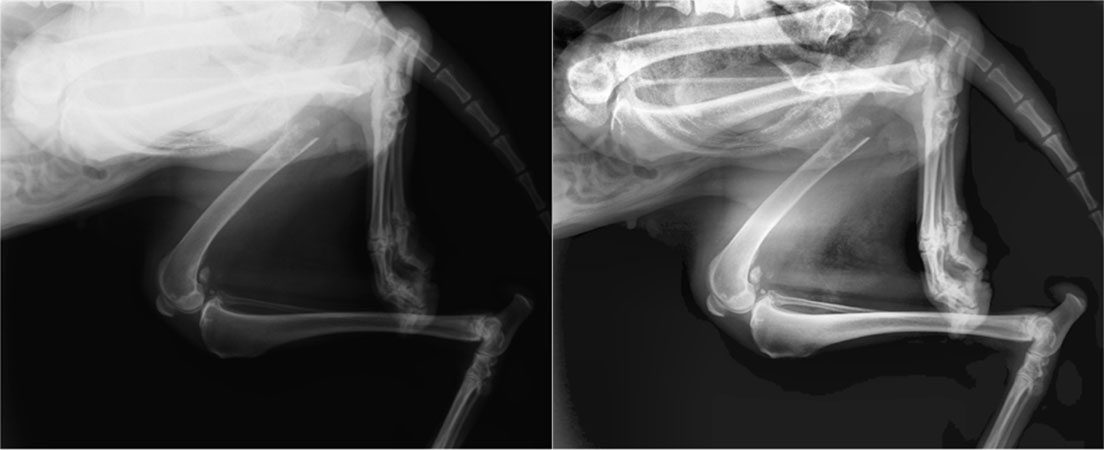
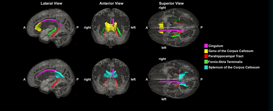
Imagen por Rayos X

El trabajo del laboratorio en el área de imagen por rayos X engloba todas las etapas de formación de imagen por rayos X desde la adquisición de los datos hasta la cuantificación de imagen pasando por calibración, pre- y post-proceso de imagen y reconstrucción tomográfica. El trabajo es este área se puede dividir en dos líneas principales: "Sistemas de rayos X clínicos" y "Sistemas de tomografía por rayos X para pequeño animal".

PET technologies

This research area is mainly focused on the development of new biomedical molecular imaging technologies based on positron emission tomography, and in its applications in clinical and preclinical research. The use of translational designs is in the centre of our activity, aiming to introduce innovative essays of new biomedical paradigms based on in vivo functional imaging.

Simulation devices

Modern teaching in Medicine and Health Sciences makes use of new tools that enable a faster and better learning, reducing risks for patients and optimizing efficiency in terms of cost. Many technologies are involved, from e-learning recources to the use of specific simulation devices which allow trainees to get acquainted with situations that are either unfrequent or highly risky for real patients.

Within this research line we investigate, in close relationship with industrial companies in this field, new technologies that may convey advantages over existing systems and devices. 

Investigación Preclínica

Molecular Imaging Probes

Molecular imaging takes advantage of the traditional diagnostic imaging techniques and introduces molecular imaging probes to determine the expression of indicative molecular markers at different stages of diseases and disorders. Molecular imaging probe is an agent used to visualize, characterize and quantify biological processes in living systems.

Neuroimaging

Schizophrenia is a chronic neuropsychiatric disorder that affects approximately 1% of the population worldwide. It is the most common chronic mental illness, representing the major economic burden on the health sector. Therefore, in recent decades, various international and national organizations have shown great concern over the impact on morbidity and quality of life. Currently, we are investigating different therapeutic strategies in the prevention of the onset of schizophrenia.

Oncology

Cancer is one of the principal leading causes of death worldwide, and thus it is one of the most studied diseases. Nuclear imaging modalities (positron emission tomography (PET) and single photon emission computed tomography (SPECT)) combined with computed tomography (CT) or magnetic resonance imaging (MRI) are the most commonly used techniques to retrieve non-invasively anatomical and functional information of tumors.

Infection and Inflammation

Early detection of unknown inflammation and nosocomial infection processes is crucial nowadays for patient’s survival. In this respect, clinical practice is taken profit of molecular imaging technologies to diagnose these pathologies in early stages. But in many cases correlation between imaging findings and patient evolution is still undetermined. Animal models are indispensable for the study of key issues in disease pathophysiology, for testing promising new treatments and also for find imaging markers of disease progression.

Quantitative Microscopy

Light microscopy employs visible light to image objects of microscopic dimensions and it is probably the most used research tool in biology.  Microscopy has historically been an observational technique. In recent years, however, the development of automated microscopes, digital sensing technologies and novel labeling probes have turned microscopy into a predominantly quantitative technique.

Cardiology

Cardiovascular diseases remain the major cause of death in the developed world. The costs generated in economic, social and human terms are immense. This has led us to propose this line to further understand the biology and basic mechanisms underlying cardiovascular diseases, identify potential novel targets, apply the information and concepts available in clinical studies of patients and, develop other biotechnological novel applications to prevent them.

Soporte a la Investigación Clínica

Intraoperative Radiation Therapy

Intraoperative Electron Radiation Therapy (IOERT) is a technique allowing for selective irradiation during cancer surgery of anatomical volumes identified as unresectable tumors, postoperative residual tumor bed or high risk tissues. It is delivered in a single dose of radiation with an electron beam. Surrounding tissues can be displaced or shielded during the procedure, allowing a more precise definition of the irradiation volume.

Image Guided Surgery

The term image guided surgery refers to those surgical procedures where the position of tracked instruments is related to preoperative or intraoperative images in order to guide the procedure. Our group has been applying these techniques to different clinical problems. The main areas of research are the following:

Imagen Óptica

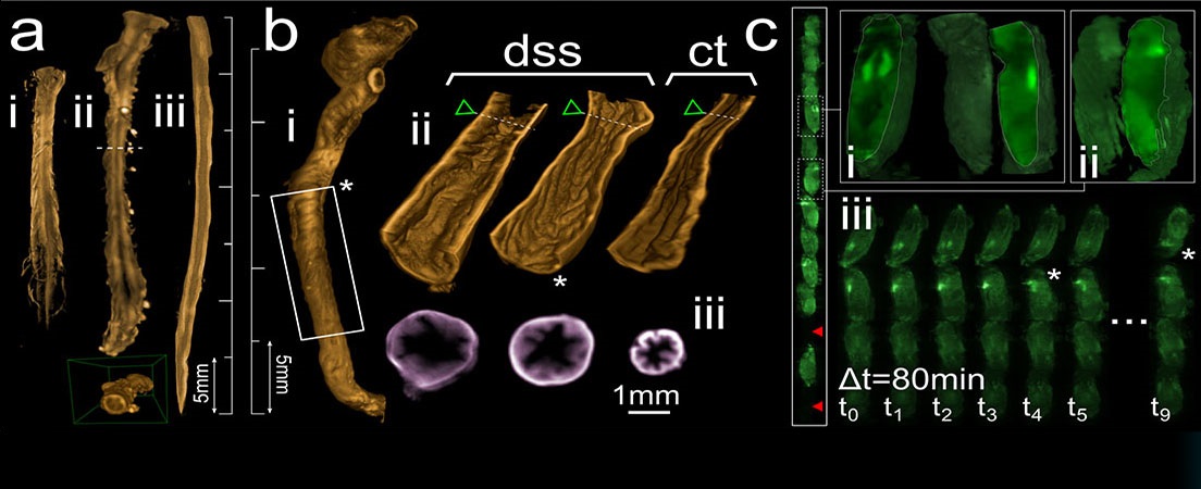
SPIM, ultramicroscopía o en general las llamadas técnicas de Haz de Láser Plano son técnicas que permiten la obtención de imágenes rápidas de alta resolución, de gran tamaño, ópticamente limpias, de especímenes tridimensionales. En el SPIM se ilumina la totalidad del órgano, muestras de tejido relativamente gruesas o cultivos organotípicos y tridimensionales, mediante un fino haz e luz que escanea a los especímenes a lo largo de un eje mientras la luz emitida es recogida a lo largo de un eje óptico perpendicular, asegurando así que el plano focal de detección coincide con el haz de luz.

Neuroimagen

El área de Neuroimagen destaca por un largo historial en el desarrollo e implementación de nuevas técnicas de adquisición y procesamiento de imágenes de Imagen por Resonancia Magnética (IRM), y de publicación en revistas de primer nivel. Forma parte y lidera la Plataforma de Neuroimagen dentro del CIBERSAM.