Unveiling the 3D cytoarchitecture of human melanome
Melanoma is the most common skin cancer. It is not deadly by its self, but can lead very easily to metastasis if it is not treated. Very little is known about the mechanisms that rule its apparition, growth and invasiveness. Conventional radiotherapy or chemotherapy, which are efficient dealing with other cancers, are useless in the case of melanoma, and the only effective approach to deal with melanoma nowadays is its surgical removal. But even after its extirpation, melanoma has a very high tendency to reappear. That is the reason why it is crucial to fully decrypt the mechanisms that rule melanoma’s behavior.
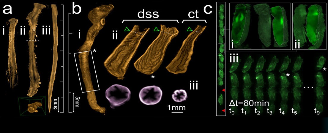
In this project 2D and 3D images of a human melanoma biopsy will be acquired using Confocal microscopy and Single Plane Illumination Microscopy (SPIM). To achieve it, melanoma samples will be cleared using different methods, such as CUBIC, and different structures will be labelled with several combinations of antibodies through a process called Immunohistochemistry.
Once the SPIM images have been acquired, typical artifacts of this microscopy modality will be corrected.


Idiomas