3D Image Quantification of Hypothalamic Neuronal Populations in Chicken Embryo Brain
The understanding of how the brain functions and develops is of great importance to the research of new advances that solve the problems caused by the numerous neurological disorders.
In the final 20% of embryonic development it is produced the transition in which the brain starts to show adult-like activity patterns. But it is unknown the exact stage in which this neuronal activation occurs. This project studies at a cellular and molecular level the last stages of chicken embryo development to define how the brain pass from being a group of neurons with spontaneous activation to be a complex organ with high neuronal activity. To achieve this objective, the project studies hypothalamic neuronal populations involved in the sleep and wake cycle as Orexin- and TH- (Tyrosine Hydroxylase) positive neurons that are positive for cFos+ protein, a neuronal activity marker.
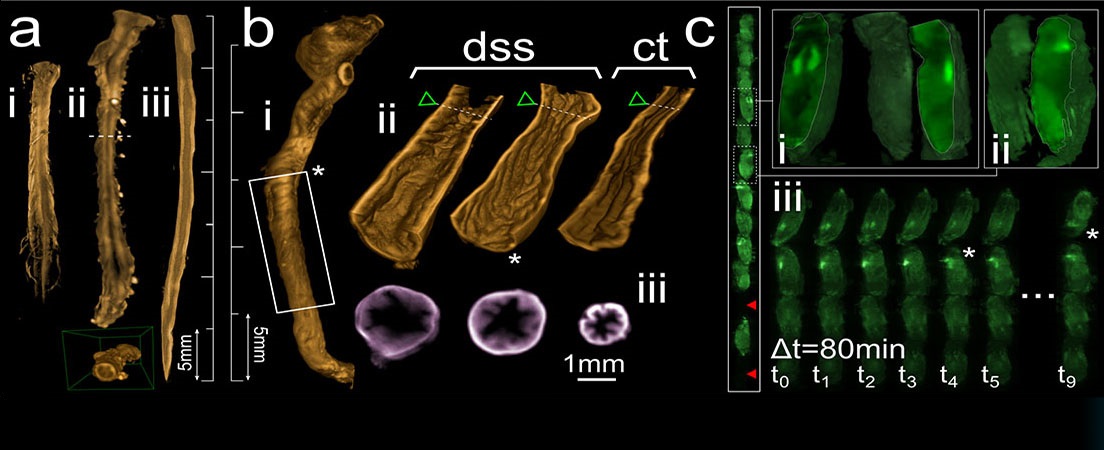
For the study of these neuronal populations, optimized tissue processing protocols must be followed. Different molecular biology methodology is used as novel tissue clearing techniques that render the samples transparent while performing the labeling of the neuronal populations under study with immunohistochemistry (IHC). Once the samples are prepared, they are imaged with new optical sectioning fluorescence microscopy such as Confocal and Single Plane Illumination Microscopy (SPIM) microscopes.
Images obtained with confocal image microscope are used to implement a 3D automatic quantification method that helps in the image analysis to achieve the final objective of the project. At the end, a combination of cellular and molecular biology, optical imaging and image processing is used in order to define the moment at which the embryo brain possesses high neuronal activity.


Idiomas