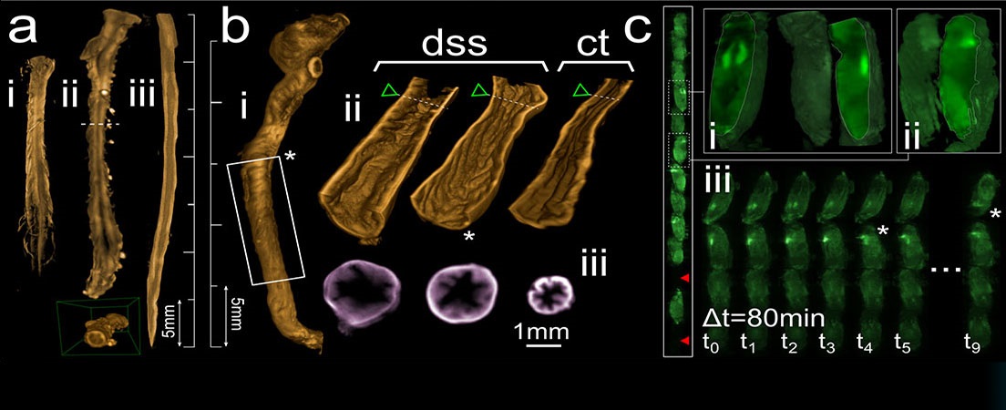
Tissue clearing methods and 3D imaging with SPIM microscopy.
Many scientific studies require three-dimensional images (3D) with cellular resolution to develop their work. An example would be the study of tumor angiogenesis. To acquire this kind of images it is needed that the tissues under study are transparent. Otherwise, light is scattered by opaque tissues and has low penetration making it impossible to take 3D images of high resolution. Tissues can be treated to become transparent using clearing agents, which turn tissues transparent, but can degrade the tissues fluorescence signal and change their size.
This report evaluates the most appropriate clearing agent for two different animal species: chick E16 embryonic brains and transgenic Drosophila melanogaster, common fruit flies expressing green fluorescent proteins (GFP).
Preliminary results from chick embryonic brains indicate that BABB and BAG successfully clear chick brains, whilst maintaining their original size. This project continued the work by studying the preservation of the fluorescence signal in cleared brains. To do so, the brains were stained firstly with antibodies and secondly with propidium iodide (PI). It was observed that none of these fluorescent compounds could penetrate more than 150-200 µm of the brains. Thus, the results of the chick embryonic brains remain as preliminary data for a future work that continues the study of the preservation of the fluorescence signal in transgenic embryos expressing endogenous fluorescence, which would not need to be stained, as they already have fluorescence signal.
In addition, Drosophila melanogaster were cleared using five compounds: BABB, BAG, FocusClear, FauxusClear and Scale. This report shows that the most appropriate clearing agent for fruit flies, taking into account the transparency of the flies, the preservation of the fluorescence and the sharpness of the images, is BABB, followed by BAG.


Idiomas