Caracterización de Microquimerismos Fetomaternales en Mus Musculus
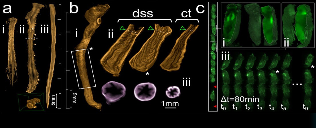
Cell traffic between mother and fetus has been well-known since the 60s. However, this bidirectional cellular exchange and the establishment of these cells in maternal tissue after birth have only been described in recent years and there are still a lot of outstanding questions. APACs (Pregnancy-Associated Progenitor Cells) or fetomaternal microchimerisms are cells that establish themselves in maternal tissues and organs for months in Mus musculus or for years in humans. These microchimerisms are able to survive and can even differentiate into cell populations that are characteristic of the tissue where they are established, modifying the tissue structure and homeostatic function of the organ. The APACs’ therapeutic potential is still a mystery although their capacity as a protective agent against neurodegenerative diseases has been described. This is why it is so important to continue researching this phenomenon and its maternal physiological effects. In the present study, several transgenic strains of Mus musculus have been used as animal models. These strains had the TdEGFP construct inserted in their genome so that different wavelengths of fluorescent light were emitted depending on the cellular lineage they belonged to. Using confocal microscopy, microchimerisms in brain, liver and heart in post-pregnant mothers have been observed. Moreover, a flow cytometer procedure has been created to determine the presence of APACs in the bloodstream before, during and after pregnancy.


Idiomas