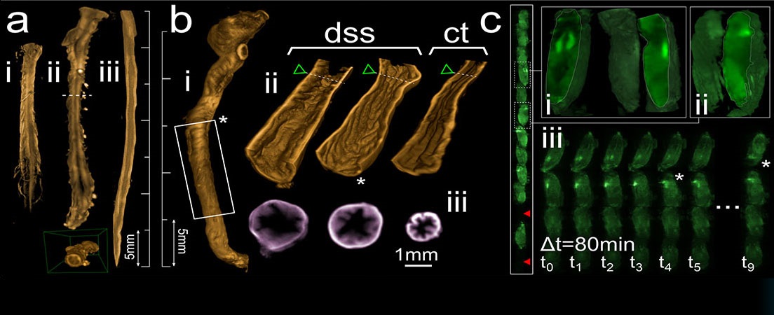
High resolution 3D-microscopy of mouse heart vasculature.
Cardiovascular diseases remain the number one cause of death globally. There is an ongoing desire to study the distribution and structural changes of the micro- and macro vasculature in the diseased heart in cardiovascular research groups all over the world. The ability to acquire high resolution 3D-images of the heart vasculature enables to study heart diseases more in detail and eventually obtain interesting new findings and new treatments. In this work we introduce a pipeline for high resolution 3D-imaging of the mouse heart vasculature with confocal- and Single Plane Illumination Microscopy (SPIM). To achieve high resolution 3D-images, protocols for optical tissue clearing (CUBIC -technique) were combined with immunohistochemical methods (IHC) and intracardial perfusion of fluorescent-labeled lectins, enabling the visualization of heart vasculature at greather depths. We here also describe the methods used for image processing of the acquired data, mainly for correction of SPIM-image artifacts and for segmentation of the structures of interest. In this work, the enhanced imaging depth and antibody penetration in cleared tissue compared with uncleared samples is assessed and quantified. SPIM bridges the gap between confocal microscopy and micro-CT and therefore is a promising relatively new imaging technique in the field of cardiovascular research.


Idiomas