Bio
Juan José Vaquero is Professor of Bioengineering at the Departamento de Bioingenieria e Ingeniería Aeroespacial, Universidad Carlos III de Madrid. He is Telecommunication Engineer from the Universidad Politécnica de Madrid (1988), and European EUR ING (1992). He also holds a Master of Bioengineering degree from the UNED and a PhD in Medical Imaging from the Universidad Politécnica de Madrid.
He was a Fogarty Fellow at the National Institutes of Health (Bethesda, Maryland, USA), where he was the principal engineer for the development of a new generation of small-animal PET systems. In 2001, he returned to Madrid and joined the Unidad de Medicina y Cirugía Experimental (Experimental Medicine and Surgery Unit) of the Hospital General Universitario Gregorio Marañón as a Ramón y Cajal Fellow in 2007, and he became a senior scientist at the Fundación para la Investigación Biomédica del Hospital Gregorio Marañón (FIBHGM), where he manages the preclinical molecular imaging activities.
His current research is focused on the development of small-animal molecular imaging systems and their associated instrumentation and data analysis.
Teaching
I teach biomedical instrumentation at the Universidad Carlos III de Madrid, Study Program Bachelor Degree in Biomedical Engineering. This degree was launched in 2010 and since then it has become one of the most pretigious bioengineering degrees in Spain. The degree is taught in English, and the number of students from abroad is increassing every year.
Selected Publications
Papers (last 10 years)
2017
- G. Konstantinou, R. Chil, M. Desco, and J. J. Vaquero, “Sub-Surface Laser Engraving Techniques for Scintillator Crystals: Methods, Applications and Advantages,” IEEE Trans. Radiat. Plasma Med. Sci., pp. 1–1, 2017, DOI: 10.1109/TRPMS.2017.2714265.
- M. V. Gómez-Gaviro et al., “Optimized CUBIC protocol for 3D imaging of chicken embryos at single-cell resolution,” Development, vol: 144, issue: 11, pp: 2092-2097, 2017.
2016
- K. M. Abushab et al., “Evaluation of PeneloPET Simulations of Biograph PET/CT Scanners,” IEEE Trans. Nucl. Sci., vol. 63, no. 3, pp. 1367–1374, Jun. 2016, DOI: 10.1109/TNS.2016.2527789.
- J. Cal-Gonzalez, M. Perez-Liva, J. L. Herraiz, J. J. Vaquero, M. Desco, and J. M. Udias, “Tissue-Dependent and Spatially-Variant Positron Range Correction in 3D PET,” IEEE Trans. Med. Imaging, vol. 35, no. 1, p. 369, Jan. 2016, DOI: 10.1109/TMI.2015.2511958.
- L. M. Fraile et al., “Experimental validation of gallium production and isotope-dependent positron range correction in PET,” Nucl. Instruments Methods Phys. Res. Sect. A Accel. Spectrometers, Detect. Assoc. Equip., vol. 814, pp. 110–116, 2016, DOI: 10.1016/j.nima.2016.01.013.
- J. L. Herraiz et al., “Automatic Cardiac Self-Gating of Small-Animal PET Data,” Mol. Imaging Biol., vol. 18, no. 1, pp. 109–116, Feb. 2016, DOI: 10.1007/s11307-015-0868-y.
- J. M. Mateos-Pérez et al., “Functional segmentation of dynamic PET studies: Open source implementation and validation of a leader-follower-based algorithm,” Comput. Biol. Med., vol. 69, pp. 181–188, Feb. 2016, DOI: 10.1016/j.compbiomed.2015.12.012.
- I. Nehrhoff et al., “3D imaging in CUBIC-cleared mouse heart tissue: going deeper,” Biomed. Opt. Express, vol. 7, no. 9, pp. 3716–3720, Sep. 2016, DOI: 10.1364/BOE.7.003716.
- S. Paonessa, S. Di Pascoli, E. Balaban, and J. J. Vaquero, “Design and Development of a Wireless Infrared EEG Recorder for chicken embryos,” 2016 Ieee Int. Symp. Med. Meas. Appl., pp. 487–492, 2016.
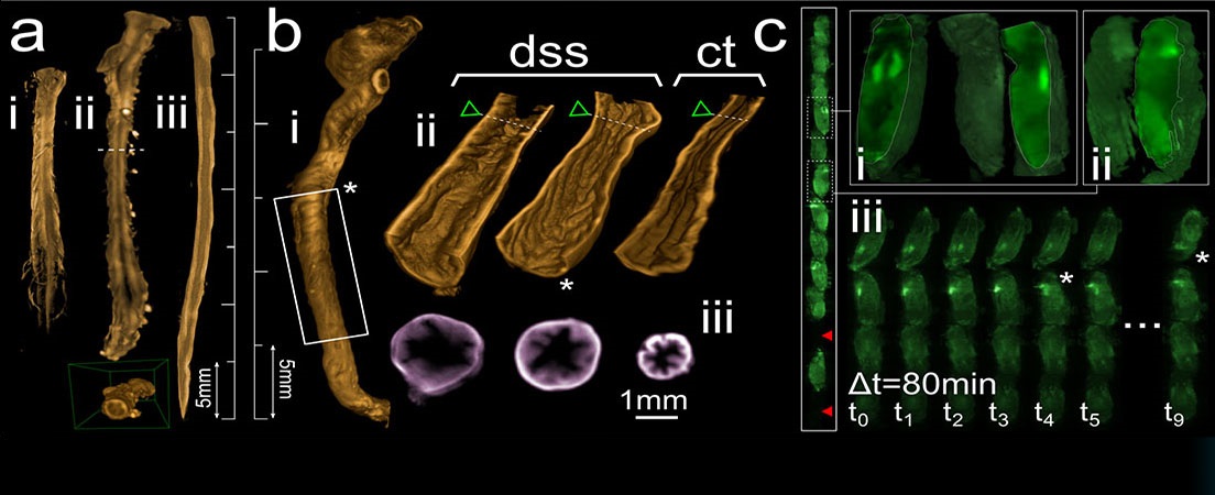
- B. Zufiria et al., “3D imaging of the cleared intact murine colon with light sheet microscopy,” in SPIE BiOS, 2016, vol. 9713, p. 97130Q, DOI: 10.1117/12.2212039.
2015
1. J.J. Vaquero and PE. Kinahan, “Positron Emission Tomography: Current Challenges and Opportunities for Technology Advances in Clinical and Pre-Clinical Imaging Systems”, Annual Review of Biomedical Engineering, Vol. 17, pp 385–414, December 2015 Q1
2. J. Cal-González, M. Pérez-Liva, J.L. Herraiz, J.J. Vaquero, M. Desco, J.M. Udías, “Tissue-Dependent and Spatially-Variant Positron Range Correction in 3D PET”, IEEE Transactions On Medical Imaging, vol. 34, no. 11, Nov 2015
3. P.A. Gómez-García, A. Arranz, M. Fresno, M. Desco, U. Mahmood, J.J. Vaquero, J. Ripoll, “Fluorescence multi-scale endoscopy and its applications in the study and diagnosis of gastro-intestinal diseases: set-up design and software implementation”, Proc. of SPIE Vol. 9531 953111
4. J.L. Herraiz, E. Herranz, J. Cal-González, J.J. Vaquero, M. Desco, L. Cussó, J.M. Udias, “Automatic Cardiac Self-Gating of Small-Animal PET Data”, Mol Imaging Biol, DOI: 10.1007/s11307-015-0868-y, 2015 Q2
5. E. Lage, V. Parot, S.C. Moore, A. Sitek, J.M. Udías, S.R. Dave, M. Park, J.J. Vaquero and J.L. Herraiz, “Recovery and normalization of triple coincidences in PET”, Medical Physics, vol. 42, no. 3, pp. 1398-1401, doi: 10.1118/1.4908226, 2015 Q2
6. J.F.P.J. Abascal, M. Abella, A. Sisniega, J.J. Vaquero and M. Desco, “Investigation of different sparsity transforms for the PICCS algorithm in small-animal respiratory gated CT”, PLoS ONE 10 (4): e0120140. doi:10.1371/journal.pone.0120140 2015 Q1
7. P.M. Gordaliza, J.M. Mateos-Pérez, P. Montesinos, J.A. Guzmán-de-Villoria, M. Desco, J.J. Vaquero, “Development and validation of an open source quantification tool for DSC-MRI studies”, Computers in Biology and Medicine, vol. 58, pp. 56–62, 2015 Q3
2014
1.- “Application of the compressed sensing technique to self-gated cardiac cine sequences in small animals”, Magn Reson Med. Aug;72(2):369-80. doi: 10.1002/mrm.24936, 2014
2.- “Comparison of methods to reduce myocardial 18F-FDG uptake in mice: calcium channel blockers versus high-fat diets”, PLoS One. 2014 Sep 19;9(9):e107999. doi: 10.1371/journal.pone.0107999, 2014
3.- “Dual-exposure technique for extending the dynamic range of x-ray flat panel detectors”, Phys. Med. Biol., vol. 59, pp. 421–439, doi: 10.1088/0031-9155/59/2/421, 2014
4.- “Modification of the TASMIP x-ray spectral model for the simulation of microfocus x-ray sources”, Med. Phys., vol. 41, pp. 011902, doi: 10.1118/1.4837220, 2014
2013
1.- ”jClustering, a n Open Framework for the Development of 4D Clustering Algorithms”, PLoS One, vol. 8, no. 8, doi: 10.1371/journal.pone.0070797, 2013
2.- “Automatic TAC extraction from dynamic cardiac PET imaging using iterative correlation from a population template” Computer Methods And Programs In Biomedicine, vol. 111, no. 2, pp.308-314, doi: 10.1016/j.cmpb.2013.04.010, 2013
3.- “MRI compatibility of position-sensitive photomultiplier depth-of-interaction PET detectors modules for in-line multimodality preclinical studies”, Nuclear Instruments and Methods in Physics Research A, vol. 702, pp. 83-87, 2013
4.- "Use of Split Bregman denoising for iterative reconstruction in fluorescence diffuse optical tomography". J Biomed Opt, 18(7): 076016 (8 pp), 2013
5.- "Positron range estimations with PeneloPET". Phys Med Biol, 58(15): 5127-5152, 2013
6.- "Monte Carlo study of the effects of system geometry and antiscatter grids on cone-beam CT scatter distributions". Med Phys, 40: 051915 (19pp), 2013
7.- "Improved dead-time correction for PET scanners: application to small-animal PET". Phys Med Biol, 58(7): 2059-2072, 2013
8.- "Massively parallelizable list-mode reconstruction using a Monte Carlo-based elliptical Gaussian model". Med Phys, 40(1): 012504 (11pp), 2013
2012
1.- “A method for small-animal PET/CT alignment calibration”, Phys. Med. Biol. 57 N199-N207–7518, 2012
2.- “Misalignments calibration in small-animal PET scanners based on rotating planar detectors and parallel-beam geometry”, Phys. Med. Biol. 57 7493–7518, 2012
3.- “Approach to Assessing Myocardial Perfusion in Rats Using Static [13N]-Ammonia Images and a Small-Animal PET”, Molecular Imaging and Biology, vol. 14, pp. 541-545, 2012
4.- “Accuracy of CT-based attenuation correction in PET/CT bone imaging”, Phys. Med. Biol., vol. 57, pp. 2477–2490, 2012
5.- “NEMA NU 4-2008 Comparison of Preclinical PET Imaging Systems”, J Nuc Med, Vol. 53, no. 8, pp. 1300-1309, 2012
6.- “Waking-like brain function in embryos”, Current Biology, vol. 22, pp. 852-861, 2012
7.- “Software architecture for multi-bed FDK-based reconstruction in X-ray CT scanners”, Computer Methods and Programs in Biomedicine, vol. 107, pp. 218-232, 2012
8.- “A method for small-animal PET/CT alignment calibration”, Phys Med Biol, Vol. 57, pp. 199–207, 2012
9.- "Influence of absorption and scattering on the quantification of fluorescence diffuse optical tomography using normalized data", J Biomed Opt 17, 036013, DOI:10.1117/1.JBO.17.3.036013, 2012
2011
1.- “GPU-Based Fast Iterative Reconstruction of Fully 3D PET Sinograms”, IEEE Transactions on Nuclear Science, vol. 58, no. 5, pp. 2257-2263, 2011
2.- “Fully 3D GPU PET reconstruction”, Nuclear Instruments and Methods in Physics Research A, vol. 648, pp. S169-S171, 2011
3.- “Study of CT-based positron range correction in high resolution 3D PET imaging”, Nuclear Instruments and Methods in Physics Research A, vol. 648, pp. S172-S175, 2011
4.- “Fluorescence diffuse optical tomography using the split Bregman method”, Med Phys 38, 6275-84, 2011
5.- "Split operator method for fluorescence diffuse optical tomography using anisotropic diffusion regularisation with prior anatomical information", Biomedical Optics Express 2(9), 2632–2648, 2011
6.- “Feasibility of U-curve method to select the regularization parameter for fluorescence diffuse optical tomography in phantom and small animal studies”, Optics Express, vol. 19, no. 12, pp. 11490-11506, 2011
7.- “Chronic Cannabinoid Administration to Periadolescent Rats Modulates the Metabolic Response to Acute Cocaine in the Adult Brain”, Molecular Imaging and Biology, vol. 13, no. 3, pp. 411-415, 2011
8.- “NEMA NU 4-2008 Performance Measurements of Two Commercial Small-Animal PET Scanners: ClearPET and rPET-1”, IEEE Transactions on Nuclear Science, vol. 58, no. 1, pp. 58-65, 2011
2010
1.- “Effects of the super bialkali photocathode on the performance characteristics of a position-sensitive depth-of-interaction PET detector module”, IEEE Trans on Nuclear Science, vol. 57, no. 5, pp. 2437-2441, 2010
2.- “A SPECT Scanner for Rodent Imaging Based on Small-Area Gamma Cameras”, IEEE Transactions on Nuclear Science, vol. 57, no. 5, pp. 2524-2531, 2010
3.- “Data acquisition electronics for gamma ray emission tomography using width-modulated leading-edge discriminators”, Phys Med Biol, 55(15): 4291-4308, 2010
4.- “Performance evaluation of SiPM photodetectors for PET imaging in the presence of magnetic fields”, Nuclear Instruments and Methods in Physics Research A, vol. 613, pp. 308–316, 2010
2009
1.- M. Abella, J.J. Vaquero, M.L. Soto-Montenegro, E. Lage, M. Desco. “Sinogram bow-tie filtering in FBP PET reconstruction”, Med Phys, 36(5): 1663-1671, 2009
2.- “Design and performance evaluation of a coplanar multimodality scanner for rodent imaging”, Phys Med Biol, 54(18): 5427-5441, 2009
3.- “The chemokine receptor CXCR4 and the metalloproteinase MT1-MMP are mutually required during melanoma metastasis to lungs”, Am J Pathol, 174(2): 602-612, 2009
4.- “Automated Method for Small-Animal PET Image Registration with Intrinsic Validation”, Molecular Imaging and Biology, vol. 11, pp. 107-113, Mar 2009
5.- “Assessment of airway distribution of transnasal solutions in mice by PET/CT imaging”, Molecular Imaging and Biology, 11(4):263-268, 2009
6.- “PeneloPET, a Monte Carlo PET simulation tool based on PENELOPE: features and validation”, Phys Med Biol, 54: 1723-1742, 2009
7.- “Detection of Visual Activation in the Rat Brain Using 2-deoxy-2-[18F]fluoro-D-glucose and Statistical Parametric Mapping (SPM)”, Molecular Imaging and Biology, vol. 11, pp. 94-99, 2009
8.- “Automatic Quantification of Histological Studies in Allergic Asthma”, Cytom Part A, 75A(3): 271-277, 2009
9.- “Research at the Medical Imaging Laboratory, CIBERSAM CB07/09/0031”, Eur J Psychiat, 23(Supl.): 43-48, 2009
2008
1.- “Real-time digital timing in positron emission tomography”, IEEE Trans Nucl Sci, vol. 55, no. 5, pp. 2531-2540, 2008
2.- “Assessment of a New High-Performance Small-Animal X-ray Tomograph”, IEEE Trans Nucl Sci, 55(3), pp. 898-905, 2008
3.- "Extraction of the respiratory signal from small-animal CT projections for a retrospective gating method". Phys Med Biol, 53, pp. 4683-4695, 2008
4.- “Augmented acquisition of cocaine self-administration and altered brain glucose metabolism in adult female but not male rats exposed to a cannabinoid agonist during adolescence”, Neuropsychopharmacol, 33(4):806-13, Mar, 2008
5.- “High resolution image in bone biology II. Review of the literature”, Med Oral Patol Oral Cir Bucal; Jan1;13(1):E31-5, 2008
Book chapters
- JJ Vaquero and SL Bacharach, "Positron Emission Tomography”, Libro: "Cardiac PET", 3rd Ed., edited by V. Dilsizian, S. Achenbach, G. Pohost, Ed.: John Wiley & Sons Limited, England, in press
- A Sisniega, M Desco, JJ Vaquero. “Design and assessment principles of semiconductor Flat-Panel detector based X-Ray micro-CT systems for small animal imaging”. Book chapter in “Integrated Microsystems and Nanotechnology: MEMs, Photonic and Biological Interfaces”, CRC Press, pp. 309-335, ISBN: 978-1-4398-3620-0, 2012
- J Lafuente, JJ Vaquero, J Sánchez. Secuencias en resonancia magnética. In: “Aprendiendo los fundamentos de la Resonancia Magnética”, E Oleaga, J Lafuente, Editorial Médica Panamericana, 2006, pp 33-43. ISBN 84-9835-121-9
- JJ Vaquero. “Medicina nuclear e Imagen Molecular”, in Ingeniería Biomédica e Imágenes Médicas, Ediciones de la Universidad Castilla-La Mancha, 2006, pp145-158, ISBN 84-8427-426-8
- JJ Vaquero. Imagen Molecular. In: Cáncer. P García-Barreno, D Espinós-Pérez and M Cascales-Angosto, Eds. Madrid: Instituto de España, 2003; pp 283-310. ISBN 84-85559-57-6
Patents in exploitation
- Planning System for Intraoperative Radiation Therapy and Method for Carrying out Said Planning, US 2011/0052036 A1, 03/03/2011
- Procedimiento y dispositivo para la detección y discriminación de eventos válidos en detectores de radiación gamma, PCT/ES2009/070456, 26/10/2009
- Incubadora para imagen con radiación no ionizante, WO 2009/077626 A1, 25/06/2009
- Multi-Modality Tomography Apparatus, US 2009/0213983 A1, 27/08/2009
- Sistema de planificación para radioterapia intraoperatoria y procedimiento para llevar a cabo dicha planificación, PCT/ES2008/000240, 14/04/2008, WO 2009/127747 A1, 22/10/2009
- Incubator for non-ionizing radiation imaging, US2011/0125010A1, 19/12/2007
- Incubadora para imagen con radiación no ionizante, PCT/ES2007/070214, 19/12/2007
- Multi-Modality Tomography Apparatus, PCT/ES2006/070150, 26/10/2006
- Tomography scanner with axially discontinuous detector array, PCT/US2004/030656, 20/09/2004
- Compact modular, configurable base for position sensitive photomultipliers, US 6,642,493 B1, 04/11/2003
- A multi-slice PET scanner from side-looking phoswich scintillators, PCT/US98/22875, WO 99/24848, 20/05/1999
Contact
Departamento de Bioingeniería e Ingeniería Aeroespacial
Universidad Carlos III de Madrid
Avda. de la Universidad, 30
28911 Leganés, Madrid. Spain
juanjose.vaquero (at) uc3m.es


Languages