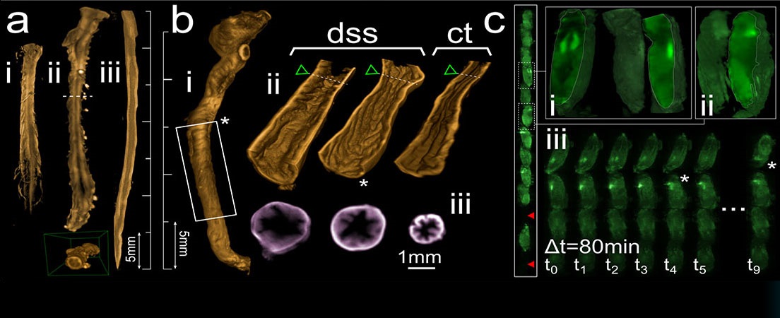
3D image of a stroke rat brain by selective plane illumination microscopy (SPIM).
The brain is the most unknown organ in the human body. Not only its complexity and its key functions for life but also brain illnesses make the brain an important organ to investigate. Very recently, new clearing protocols as CUBIC have been described to clear whole mouse organs such as brain. This method allows us to visualize in 3D whole brain by using a SPIM microscope (selective plane illumination microscopy). In neurovascular diseases such stroke, the brain suffers structural and functional changes that can be visualized with this methodology.
The present work aims to optimize the CUBIC protocol to clear rat brains and visualize the damaged area in rats with stroke using SPIM microscopy. To carry out it, this rat brains were cleared and stained with CUBIC protocol. Together with the CUBIC protocol, immunohistochemistry assays were performed with Sox-2, RecA and caspase-3 antibodies to stain progenitor/-stem cells, blood vessels and apoptosis cells respectively.
Our results suggest that the CUBIC protocol is appropriated to clear rat brain but needs some future work to clear the deepest areas and achieve a better antibody penetration through the tissue. SPIM microscopy demonstrates its efficacy showing a 3D brain structures immunostained. Understanding the 3D structure of the brain and the capability to visualize cells localization inside the whole brain will be very useful to unveil new therapeutic targets for pathologies such as stroke.


Idiomas