3D imaging of human cleared melanoma with spim microscopy
Melanoma is one of the most common types of cancer and is characterized by its aggressiveness and tendency to cause metastasis even after removal of the primary tumour, with a survival rate of only 24% after it takes place. The high level of angiogenesis showcased by all cancerous processes and particularly melanoma plays a significant role in this process; nevertheless, we lack a detailed model of the architecture of this cancer-induced new vasculature and the possible information it would provide for its palliation and prevention.
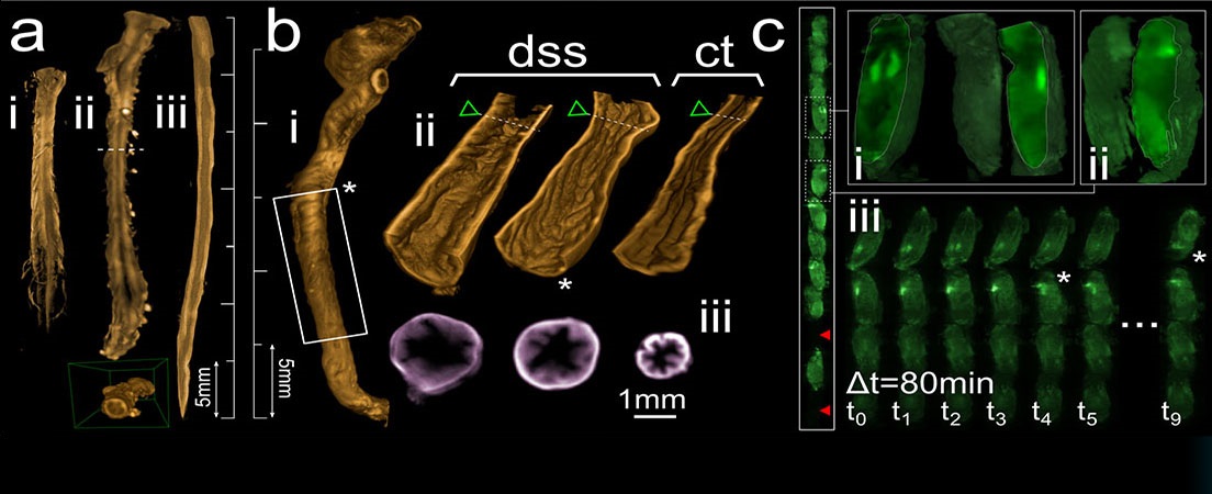
Single Plane Illumination Microscopy (SPIM) can be used to generate high resolution 3D images of tissue samples while preserving their original structure intact and thus the relevance of said data for clinical applications. SPIM imaging is performed in combination with the CUBIC (Clear, Unobstructed Brain Imaging Cocktails) optical tissue clearing protocol and immunohistochemistry (IHC) vasculature labelling methods to study human melanoma samples and obtain a 3D reconstruction of its structure.
This project focused on the optimization of the CUBIC protocol coupled with IHC for effective use on human melanoma samples for the generation of truthful 3D models of its vasculature. The clearing protocol details and lengths were adjusted for maximum efficiency with the best possible result, while the
specifics of the required image acquisition and processing steps were assessed in their difficulty and feasibility. In the end, high-quality 3D images of human melanoma vasculature were successfully generated and visualized, paving the way for further future studies.


Languages