Stratification of abnormal density associated with tuberculosis
Tuberculosis (TB) is an infectious disease caused by the bacillus Mycobacterium tuberculosis. It has existed for miles of years and remains a major global health problem, being the main cause of death from an infectious agent for the past five years.
Certain drugs sometimes succeed to cure infected people. However, recent studies by the World Health Organization (WHO) sustain that the number of people developing Multi-Drug Resistance (MDR) is increasing. Meaning that the current drugs are starting to be nonfunctional, and a better understanding of the diseased is needed.
Diagnosis is a crucial step. On occasion, the infection is difficult to detect micro-CT at first glance by the doctor because it could be in a latent state. Nevertheless, nowadays a complete sequence of steps exists, that ensures the diagnosis of the bacillus. The main three stages involved are chest radiography, Human Immunodeficiency Virus (HIV) test and sputum smear and culture.
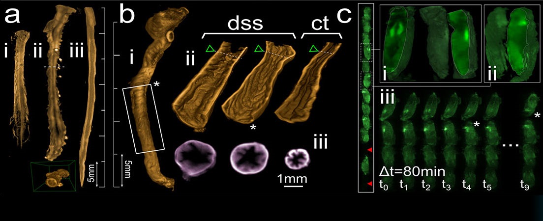
Imaging plays a crucial role in the diagnosis and management of tuberculosis. Medical imaging in the field of TB has been developed and new modalities have shown good results (Computed Tomography, Magnetic Resonance, Positron Emission Tomography and Single-photon Emission Tomography). However, Computed Tomography (CT) has the gold standard regarding the good resolution of the images for the lesion detection.
CT images have been demonstrated to be good support for the analysis of the evolution of TB. It also provides metrics that allow other image modalities to complement the information it contains. One example is histopathological images. They have more accurate information of the distribution of the disease in the tissue, therefore it is a good idea to extrapolate that information into CT images as this could contribute to a more accurate diagnosis from the imaging modalities.


Languages