High Resolution 3D Microscopy of Developmental Neurogenesis in Chick Embryo Models.
It is unknown when and how the brain makes the developmental transition from a group of cells that are activated spontaneously to a complex organ that functions on a large scale, such as the brain in higher vertebrate animals. The comprehension of how the brain develops and functions at a cellular and molecular level is of fundamental importance in the search for answers to a broad range of questions relating to neurological disorders.
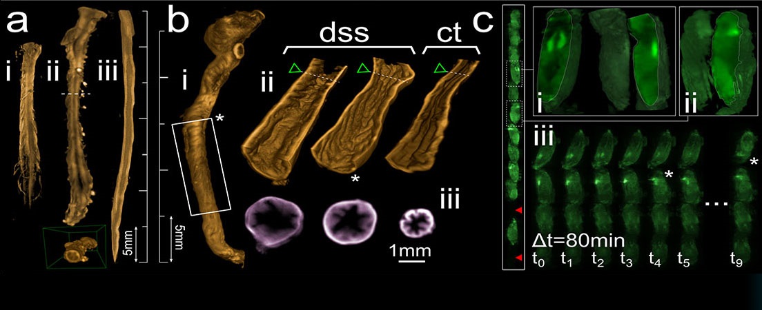
In the final 30% of embryonic life of higher vertebrates, brains start to show adult-like forms of activity. The hypothalamus area in the brain is one of the regions involved in regulating the different stages of sleep and waking. This project develops and optimizes the methodology to study hypothalamic Orexinand TH- (Tyrosine Hydroxylase) positive neurons, involved in sleep and waking, that are also positive for cFos+ protein, a marker of neuronal activity. Chick embryo brains during the last 30% of embryonic life will be studied using novel fluorescence imaging techniques. Modern optical clearing methods make it possible to render the samples transparent while performing immunohistochemistry (IHC). Visualization of positive neuronal populations in these large, cleared samples has been carried out using optical sectioning fluorescence microscopy, such as Confocal and Single Plane Illumination Microscopy (SPIM) microscopes.


Languages